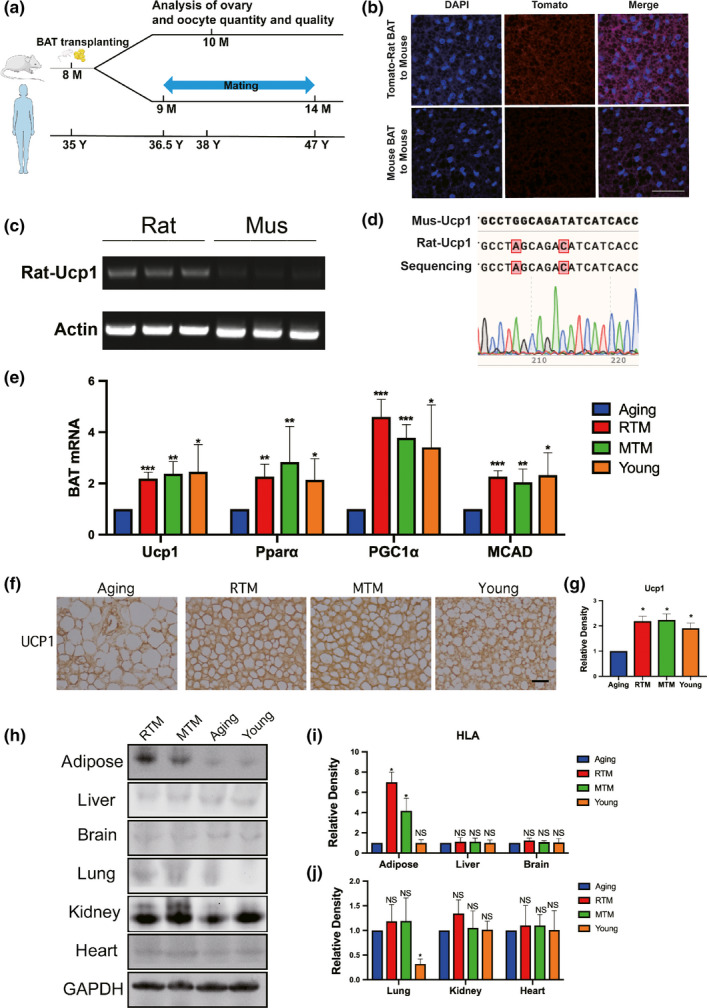
Figure 1.

Rat‐to‐mouse (RTM) xenotransplanted brown adipose tissue (BAT) was functional well and did not cause injurious histocompatibility in aging mice. (a) Scheme of experimental design. Eight‐month‐old aging mice were divided into four groups, RTM and MTM groups received brown adipose tissue (BAT) from 1‐month‐old rats or mice, then recovered for 1 month. Aging mice accepted a pseudo operation (cut and stitch up without BAT transplantation). At 9 months old, mating assays started, five mice for each group. At about 10 months old, when the first litter is delivered, more mice were used for all other experiments. The age line of human corresponds to age in mice. (b–d) Verification of xenotransplanted rat BAT 3 weeks after xenotransplantation. (b) BAT paraffin sections were stained with DAPI (blue) and fluorescence image showed that tomato fluorescence (red) was very remarkable in the RTM group while very low in the MTM group. (c) RT–PCR with rat‐specific UCP1 primers detects a bright band with expected size from all three RTM mice, while no observable bands from three MTM mice. (d) The bright band from (c) was identified to be rat UCP1 by Sanger sequencing. (e) qPCR showed that the mRNA levels of four BAT marker genes, UCP1, PPARα, PGC‐1α, and MCAD, were significantly rescued close to the young group. (f) UCP1 immunohistochemistry in BAT paraffin section showed that UCP‐1 level in RTM and MTM group, and young group were significantly higher than in the aging group. (g) Quantification of (f). (h) HLA‐A blot showed that HLA‐A level in BAT of RTM and MTM groups was significantly higher than aging or young group; HLA‐A level in the lung of young group is significantly low; HLA‐A levels in other main tissues (liver, spleen, brain, kidney, heart) are similar among all four groups. (i,j) Quantification of HLA‐A levels in (g). Scale bar, 20 µm. *p < 0.05, **p < 0.01, ***p < 0.001 are considered significantly different
