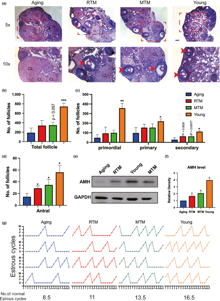
Figure 3.

RTM significantly improved the ovarian performance of aging mice. (a–d) Hematoxylin–eosin (HE) staining of continuous ovary paraffin sections (a) and number of ovarian follicles (b–d). (b) for total follicles, (c) for primordial (left), primary (middle), and secondary (right) follicles. (d) for antral follicles. There was no significant difference for the numbers of primordial, primary, secondary, and total follicles between RTM and aging groups, but the number of antral follicles in RTM and MTM groups was a fold more than the aging group. For better vision in (a), a subregion (dot‐line rectangle region) of the upper image (5×) in each group was magnified and put at the lower panel (10×). CL, corps lutein. Antral follicles were arrow‐pointed. (e) Blot showed that AMH levels in RTM, MTM, and young groups were significantly higher than in the aging group. (f) Quantification of AMH levels of ovaries from four groups. (g) Plots of estrus cycle curves within 21 test days, four mice were used for each group. There are more cycles in RTM, MTM, and young groups than in the aging group. Total numbers of estrus cycle from four groups were shown at the bottom of each plot; Scale bar, 200 µm. *p < 0.05, **p < 0.01, ***p < 0.001 are considered significantly different
