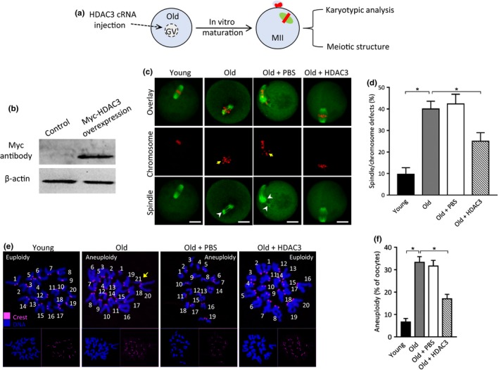
Figure 2.

HDAC3 overexpression ameliorates the maternal age‐associated meiotic defects in mouse oocytes. (a) Schematic illustration of the HDAC3 overexpression experiments. (b) Western blotting shows the efficiently overexpressed exogenous HDAC3 protein, probing with anti‐Myc antibody. (c) Young, old, old + PBS, old + HDAC3 oocytes were stained with α‐tubulin antibody to visualize spindle (green) and counterstained with PI to visualize chromosome (red). Arrows point to the misaligned chromosomes and arrowheads indicate the disorganized spindle. Scale bars: 20 µm. (d) Quantification of young (n = 116), old (n = 110), old + PBS (n = 102), old + HDAC3 (n = 108) oocytes with spindle/chromosome defects. (e) Chromosome spread of young, old, old + PBS, old + HDAC3 MII oocytes. Chromosomes were stained with Hoechst 33,342 (blue), and kinetochores were labeled with CREST (purple). Arrow points to the extra chromosome in old oocytes. (f) Histogram showing the incidence of aneuploidy in young (n = 26), old (n = 32), old + PBS (n = 30), old + HDAC3 (n = 28) oocytes. Error bars indicate ± SD. *p < .05 versus controls
