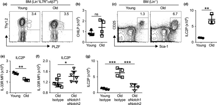
Figure 1.

Aging enhances early ILC2 development in the bone marrow. (a) Representative flow cytometry profile of common helper innate lymphoid cells (CHILP, Lin‒IL7R+α4β7+Thy1+PLZF+) in the bone marrow of young (2–3 months) and old (19–24 months) mice. Plots were pregated on Lin‒IL7R+α4β7+ cells. (b) Number of CHILP from bone marrow of young and old mice. (c) Representative flow cytometry profile of ILC2P (Lin‒CD25+Sca‐1+) from the bone marrow of young and old mice. Plots were pregated on Lin‒ cells. (d) Number of ILC2P cell from bone marrow of young and old mice. (e) Mean fluorescence intensity (MFI) of IL‐33R of ILC2P cells. (f) MFI of IL‐33R from old mice treated with isotype controls or αNotch1 and αNotch2 antibodies. (g) Number of ILC2P from young and old mice treated with isotype controls or αNotch1 and αNotch2 for 3 days. Data are from 3 to 6 mice per group; *, p < 0.05; **, p < 0.01; ***, p < 0.001
