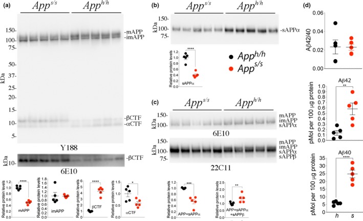
Figure 2.

Increased processing by β‐secretase and decreased processing by α‐secretase of APPSw. To test whether the App s/s rats present the expected changes in APP metabolism, we analyzed brain samples isolated from 21‐day‐old App h/h and App s/s rats, 2 females and 3 males for each genotype. (a) Western blot (WB) of postnuclear supernatant from App s/s and App h/h rats with Y188 and 6E10 (bottom). (b) WB analysis of soluble brain fractions with an ant‐sAPPα antibody showed that levels of sAPPα are significantly lower in App s/s compared to App h/h brains. (c) Because sAPPβh and sAPPβSw cannot be detected by the same antibody (see Figures 3a,b and 4d,e), to compare sAPPβh and sAPPβSw amounts, we used 6E10 and 22C11. Signal intensity was quantified with Image Lab software (Bio‐Rad). Quantification is shown on the right. (d) Aβ is produced by γ‐cleavage of βCTF, which is increased in App s/s brains. ELISA measurements of endogenous Aβ40 and Aβ42 in brain homogenates of 21‐day‐old animals. Levels of steady‐state endogenous Aβ40 and Aβ42 are increased in App s/s brains as compared to App h/h brains. The Aβ42/Aβ40 ratio did not change. These ELISA kits are specific for human Aβ40‐42, as attested by the fact that they gave no signal on brain homogenates from App w/w (expressing rat Aβ) and App δ7/δ7 (expressing no Aβ) rats (not shown). Data are represented as mean ± SEM. Data were analyzed by Student's t test and shown as average ± SEM. **p < .01; ****p < .0001
