
Figure 6.
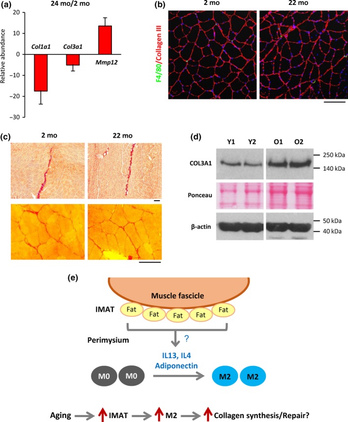
Collagen mRNA abundance, but not total collagen levels, declines in aged SKM. (a) RT‐qPCR analysis revealed markedly reduced levels of Col1a1 and Col3a1 mRNAs in old mouse SKM; by contrast, Mmp12 mRNA was strongly upregulated. (b) Immunostaining showed comparable intensity of collagen III signals in young and aged mouse skeletal muscle. (c) Picrosirius red staining showed comparable collagen deposits (red) in perimysium (upper panels) and endomysium (lower panels) areas of young and old mouse SKM. (d) Western blot analysis of collagen 3 protein levels in young and old mouse SKM. (e) Model illustrating the hypothesis of macrophage polarization in SKM. Based on our results, we propose that adipocytes in the perimysium secrete cytokines that provoke M2 polarization of macrophages. In aged skeletal muscle, IMAT increases, leading to elevated cytokine secretion and a rise in M2 macrophages. Increased M2 macrophages in turn contribute to muscle tissue repair and muscle fibrosis in aged skeletal muscle likely by promoting collagen synthesis. Scale bars in (b) and (c) represent 100 µm