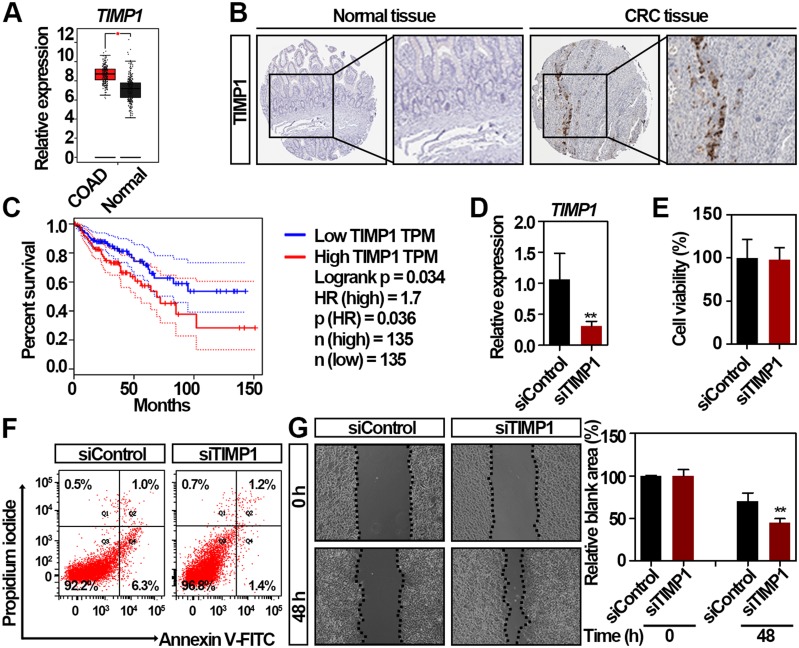
Figure 3.
Identification of TIMP1 as a potential biomarker for the worse prognosis of patients with CRC. (A) TIMP1 is significantly upregulated in CRC tissues compared with normal colorectal tissues by GEPIA. *significantly different from normal tissues; *p < 0.05. (B) The expression of TIMP1 in the CRC and non-malignant colorectal tissues. Representative immunohistochemistry images were retrieved from the Human Protein Atlas online database. Magnification, x40. (C) Survival analysis of the correlation between TIMP1 expression and overall survival in patients with CRC generated from GEPIA. Log rank tests were employed to determine the statistical significance. (D) Relative expression of TIMP1 in RKO cells after transfection with 100 nmol/L TIMP1 siRNA (siTIMP1) and control siRNA (siControl) for 48 h as assessed by qRT-PCR. Data are shown as mean ± SD. **p < 0.01. (E) Cell proliferation analysis of RKO cells after transfection with 100 nmol/L siTIMP1 and siControl for 48 h. Data are shown as mean ± SD. Two independent experiments were performed. (F) Flow cytometric analysis of Annexin V/PI staining in RKO cells after transfection with 100 nmol/L siTIMP1 and siControl for 48 h. (G) RKO cells exhibited increased cell migration after transfection with 100 nmol/L siTIMP1 and siControl as assessed by the scratching wound-healing assay. Data are shown as mean ± SD. **p < 0.01. Two independent experiments were performed. Unpaired Student’s t-test was performed (D, E, and G).

