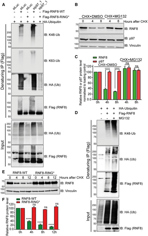
Figure 2. Homeostasis of RNF8 is controlled by auto‐ubiquitination and the ubiquitin–proteasome system.

- Western blot analysis of Flag‐RNF8 denaturing‐IP in HEK293 cells showing the ubiquitination pattern of RNF8‐WT and RNF8‐RING* variant, under siRNA‐mediated luciferase (siLuc) or p97‐depleted conditions (sip97).
- Western blot analysis of CHX chase kinetics in HeLa cells showing the degradation kinetics of RNF8 and inhibition of RNF8 degradation by simultaneous proteasome inhibition (MG132, 10 μM).
- Graph represents the quantifications of (B) (ns: not significant, P > 0.05, ****P < 0.0001; two‐way ANOVA, n = 2, mean + SEM).
- Western blot analysis of Flag‐RNF8 denaturing‐IP in HEK293 cells showing hyper‐ubiquitination of RNF8 after proteasome inhibition (MG132, 10 μM for 6 h).
- Western blot analysis of CHX chase kinetics in U2OS cells, comparing the degradation rate of Flag‐RNF8‐WT and Flag‐RNF8‐RING*. Endogenous RNF8 was depleted by shRNF8 targeting only endogenous RNF8.
- Graph represents the quantifications of (E) (ns P > 0.05, ****P < 0.0001; two‐way ANOVA, n = 3, mean + SEM).
Source data are available online for this figure.
