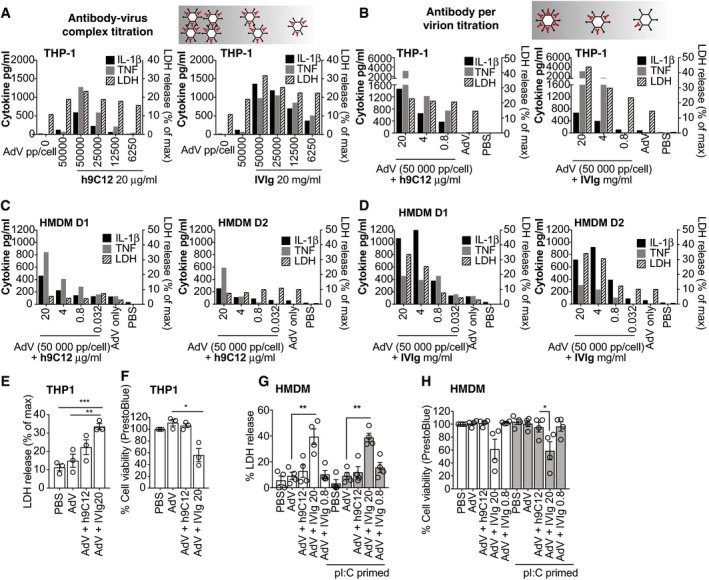
IL‐1 β, TNF (left axis) and LDH release (right axis) in the cell supernatants were measured 16 h after stimulation.
-
A
AdV‐GFP was incubated with 20 μg/ml h9C12 antibody or 20 mg/ml IVIg in PBS. These AdV‐Ab complexes were then diluted 1:2 to give final doses of viral particles being added to WT THP‐1 cells (with antibody complexed) as indicated. A single experiment representative of three independent experiments is shown.
-
B
THP‐1s were stimulated with a constant dose of AdV‐GFP (50,000 pp/cell) and complexed with decreasing antibody concentrations as indicated. A single experiment representative of two independent experiments is shown.
-
C, D
HMDM were stimulated directly (TNF) or primed (IL‐1β, LDH) with AdV‐Ab complexes as in (B). Data for each individual donor (two total) are shown.
-
E, F
THP‐1s were stimulated with AdV (50,000 pp/cell) complexed with h9C12 (20 μg/ml) or IVIg (20 or 0.8 mg/ml) for 16 h, and cell death was measured by LDH release (E) or cell viability measured by PrestoBlue assay (F) (n = 3, mean ± s.e.m. *P ≤ 0.05, **P ≤ 0.005, ***P ≤ 0.001 unpaired, two‐tailed t‐test).
-
G, H
HMDM were primed, or not, and stimulated with AdV (50,000 pp/cell) complexed with h9C12 (20 μg/ml) or IVIg (20 or 0.8 mg/ml) for 16 h, and cell death was measured by LDH release (G) or cell viability measured by PrestoBlue assay (H) (n = 4, mean ± s.e.m. *P ≤ 0.05, **P ≤ 0.005, paired, two‐tailed t‐test).

