FIGURE 4.
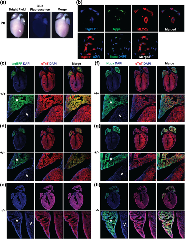
Nppa-tagBFP knock-in reporter expression in the neonatal heart. (a) Whole mount fluorescent image of Nppa-tagBFP knock-in reporter mice at P0. (b) Co-expression of Nppa-tagBFP reporter with endogenous Nppa and MLC-2a in neonatal atrial cardiomyocytes. Neonatal atrial cardiomyocytes were isolated from Nppa-tagBFP reporter knock-in mice. Isolated neonatal atrial cardiomyocytes were immunostained for MLC-2a and Nppa. tagBFP fluorescence was visualized without immunostaining. The frozen heart sections of homozygous and heterozygous Nppa-tagBFP knock-in mice, and wild type littermates at P0 were immunostained with tagBFP (c–e) or Nppa (f–h), and cTnT (c–h). The top panels show whole hearts demonstrating four chambers. The bottom panels are magnified views showing both the atrium and ventricle. A: atrium; V: ventricle

