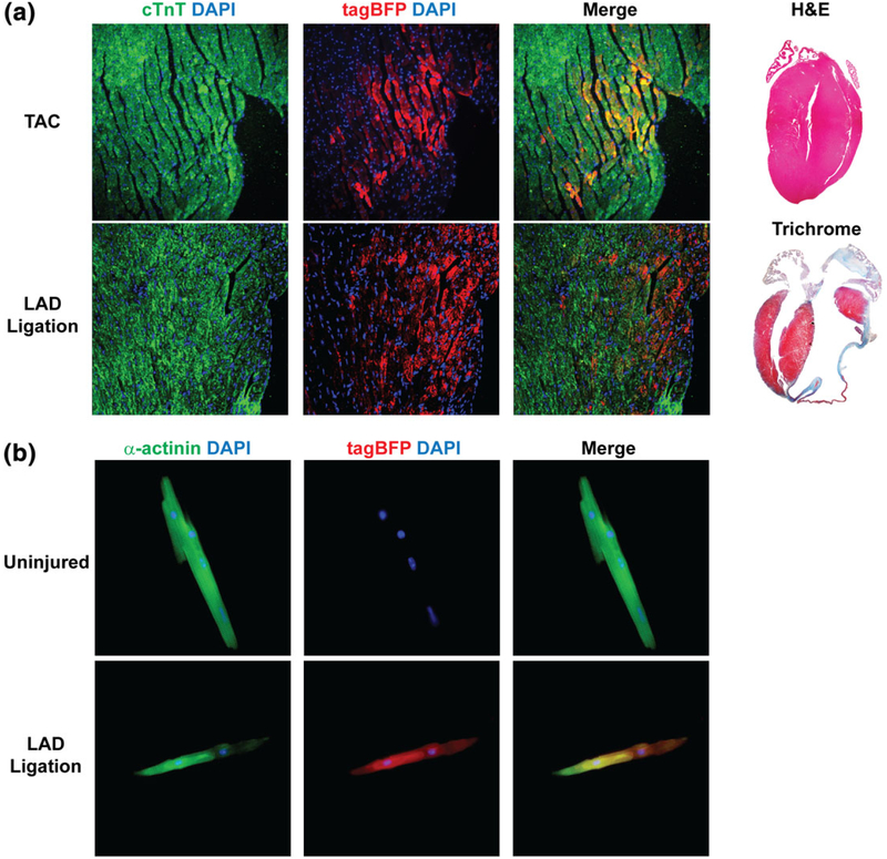
FIGURE 6.
Nppa-tagBFP knock-in reporter expression during heart diseases. (a) Immunofluorescent staining of frozen heart sections of Nppa-tagBFP knock-in reporter mice following injuries. Two months after TAC (top) or LAD ligation (bottom), heart sections of Nppa-tagBFP knock-in reporter mice were obtained from a mid-portion of septum (TAC) and a remote area of infarction (LAD ligation). Heart sections were processed and immunostained for tagBFP and cTnT. Nppa-tagBFP knock-in reporter expression in ventricular cardiomyocytes was shown. H&E (for TAC) and Trichrome (for LAD ligation) stainings were performed to demonstrate the extent of insult (right). (b) Immunofluorescent staining of isolated adult ventricular cardiomyocytes from Nppa-tagBFP knock-in reporter mice with or without injury. Three weeks after LAD ligation, ventricular cardiomyocytes were isolated from the mice using Langendorff perfusion and immunostained for tagBFP and α-actinin. Age-matched uninjured mice were used as a control

