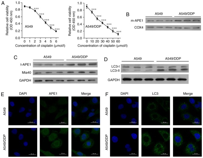
Figure 1.
Cisplatin-resistant A549 cells exhibit high levels of APE1 and autophagy. (A) A549 cells were treated with 0, 1, 2, 3, 4, 5 and 6 µmol/l cisplatin for 24 h, and the A549/DDP cells were treated with 0, 10, 20, 30, 40, 50 and 60 µmol/l cisplatin for 24 h. Cell viability was assessed by the CCK-8 assay. (B) Western blot analysis was performed to analyze the levels of mitochondrial APE1 (m-APE1) protein in A549/DDP and A549 cells. COX4 was used as loading control for the mitochondrial APE1 protein. (C) Western blot analysis of the total APE1 (t-APE1) and Mia40 protein levels in A549/DDP and A549 cells. GAPDH was used as a loading control. (D) Western blot analysis of the LC3 protein levels in A549/DDP and A549 cells. (E and F) Immunofluorescence assays were performed to assess the total APE1 and total LC3 levels in A549 and A549/DDP cells. Data represent results obtained from three independent experiments (mean ± SEM of triplicate samples). ***P<0.001, vs. 0 µmol/l cisplatin-treated A549 or A549/DDP cells. APE1, apurinic/apyrimidinic endonuclease 1; LC3, microtubule-associated protein 1A/1B-light chain 3.

