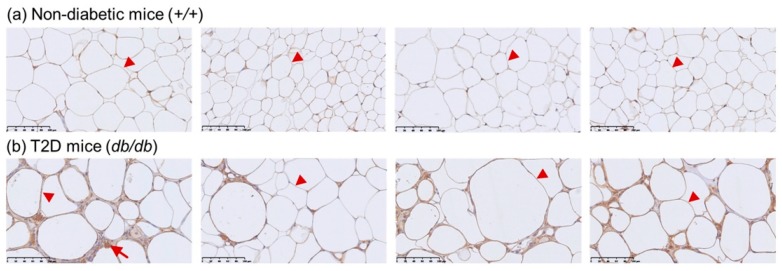
Figure 2.
Representative images of immunohistochemical staining of CETP expression in mouse adipose tissue at 32 weeks of age (magnification 400×) (a) Non-diabetic mice (the arrowhead indicates adipocytes). (b) CETP is prominently expressed in the membrane of adipocyte in the T2D mouse model (arrowhead indicates adipocyte; arrow indicates cell with signs of inflammation).

