Figure 2.
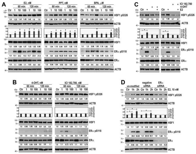
ERα is indispensable for HSF1 phosphorylation on S326 in MCF7 cells. (A) An influence of ERα agonists (E2, PPT, BPA) and (B) E2 antagonists (4-OHT and ICI 182,780) on HSF1 phosphorylation. (C) Effect of E2 on HSF1 phosphorylation after pretreatment of cells with 4-OHT or ICI 182,780 (added at concentration 100 nM for one hour before treatment with 10 nM E2 for one hour or two hours, respectively). (D) Effect of E2 on HSF1 phosphorylation in cells with down-regulated ERα. ERα phosphorylation on S118 was used as a marker of ERα activation, and ACTB was used as a protein loading control. The results of densitometric analyses are shown in the charts. An asterisk indicates a significant difference (p < 0.05) from the control (Ctr) value. Numbers below blots represent protein bands’ intensity ratios to adequate controls after normalization against ACTB.

