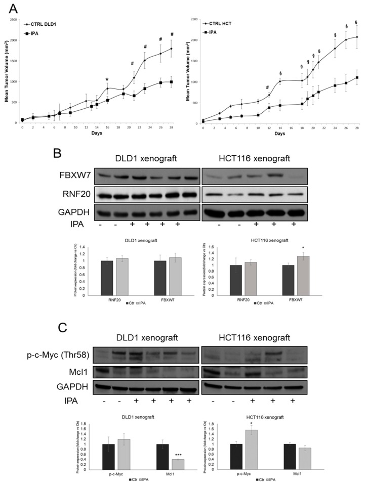
Figure 6.
IPA reduces CRC growth in vivo. (A) Tumor volume growth curve of DLD1-xenograft (left panel, n = 20 mice, 10 mice in each group) and HCT116-xenograft (right panel; n = 20 mice, 10 mice in each group) after peri-tumoral injection of IPA. Growth retardation by the compound was statistically significant for all time points labelled with * (one-way ANOVA p < 0.05), with # (one-way ANOVA p < 0.01) and § (one-way ANOVA p < 0.001). Western blot and densitometry analysis of FBXW7, RNF20 (B), phosphorylated-c-Myc (Thr58) and Mcl1 (C) in whole lysate from representative resected tumor tissues. GAPDH was used as loading control. Data are expressed as mean ± SD of at least four independent experiments. * p < 0.05, *** p < 0.005 vs. control group.

