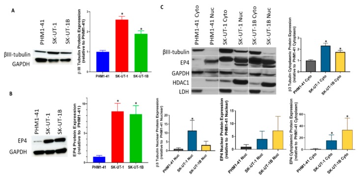
Figure 4.
Protein expression in cell lines. (A) Class III β-tubulin and (B) EP4 protein expression were analyzed via Western blot in sarcoma (SK-UT-1) and carcinoma (SK-UT-1B) cell lines relative to normal immortalized uterine myometrial cell line PHM1-41. Both class III β-tubulin and EP4 were found to be overexpressed compared to PHM1-41. The bar graphs on the left indicate densitometry measurement of the Western blots. Protein expression analysis of the nuclear and cytoplasmic fractions is shown in (C). The bar graphs below on the right indicate densitometric measurement of cytoplasmic and nuclear class III β-tubulin or EP4 proteins normalized to GAPDH. Both class III β-tubulin and EP4 proteins in the cytoplasmic fractions of SK-UT-1 and SK-UT-1B had significantly more expression compared to PHM1-41. Only class III β-tubulin in the nuclear fraction of SK-UT-1 had significantly increased expression compared to PHM1-41. The following antibodies were used as loading controls: GAPDH (total protein), histone deacetylase 1 (HDAC1) (nuclear loading control), and lactate dehydrogenase (LDH) (cytoplasmic loading control). * p < 0.01.

