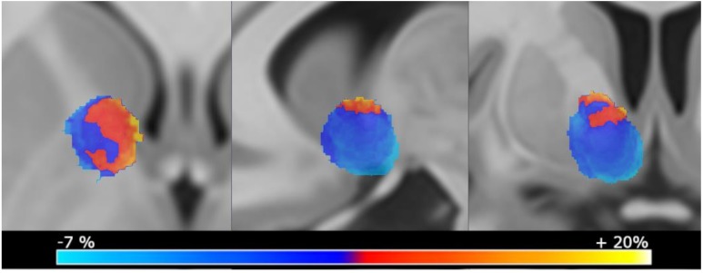
Figure 1.
Averaged weight change maps displaying mean weight changes per voxel. Only voxels that were stimulated by at least 10% of the patients were selected to control for outliers. Volumes of activated tissue (VTA) that were located more medially and apically were associated with more weight gain after intervention.

