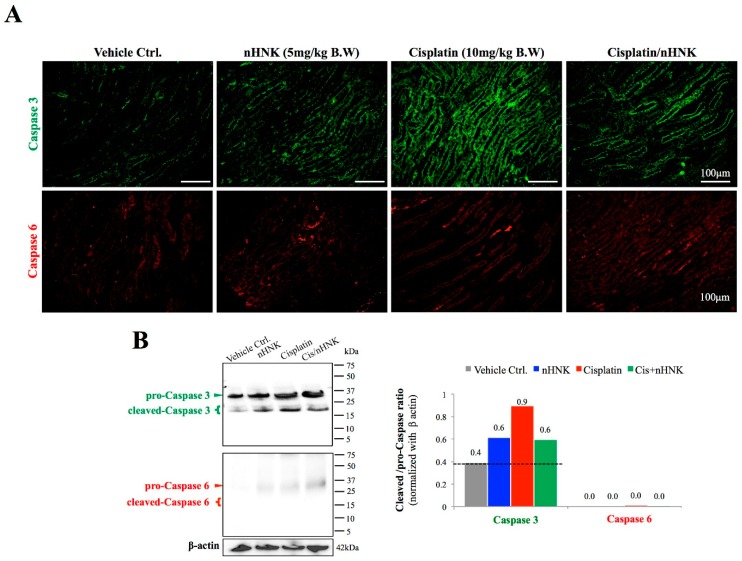
Figure 5.
Evaluation of caspase-dependent signaling pathway in cisplatin-injured animals. Caspase 3 and caspase 6 were used to evaluate the activation of cellular apoptosis process. (A) A minimal signal on both caspase 3 and caspase 6 was observed in control and nHNK alone groups. An intense caspase 3, but not caspase 6, signal was detected at the collecting duct of cisplatin-injured kidneys, which indicated that cisplatin induced pronounced cellular apoptosis in kidney collecting ducts. A decreased signal on caspase 3 was detected when nHNK was given to cisplatin-injured animals, which demonstrated that nHNK mitigated cisplatin-induced cellular apoptosis. (B) Western-blotting analysis showed cisplatin-induced cleavage of pro-caspase 3 enzyme, but these increases were attenuated when nHNK was present. Images presented are representative images of each group.

