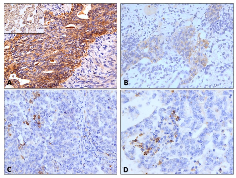
Figure 2.
Illustrative examples of CTLA-4 staining in testicular germ cell tumors. (A) and (B) CTLA-4 strong (A) and moderate (B) intensity staining in tumor cells of residual mature teratomas in lymph node metastasis and in a primary tumor (immature teratoma component of a mixed tumor) (200×). Notice the predominantly cytoplasmic but also slight membrane staining. Inset: positive control (placenta) for CTLA-4 staining, included in all slides; (C) and (D) CTLA-4 staining in scattered immune cells populating two pure embryonal carcinomas (400×). Notice the punctate, granular staining, like for PD-L1.

