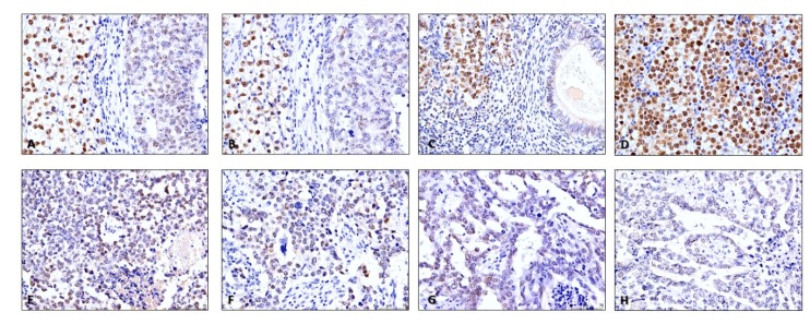
Figure 7.
Illustrative examples of mismatch repair proteins staining in testicular germ cell tumors. (A) and (B)—MLH1 (A) and PMS2 (B) staining patterns in a mixed tumor composed of seminoma (on the left) and embryonal carcinoma (on the right). Notice the clearly weaker intensity staining in embryonal carcinoma and reduced number of stained cells when compared to the seminoma component (200×); C—MSH2 staining in a mixed tumor composed of seminoma (on the left) and teratoma (on the right). Notice the clearly stronger intensity staining in seminoma when compared to the teratoma element (200×); D—MLH1 strong and diffuse intensity staining in a pure seminoma. This was the most common pattern witnessed in pure seminomas (200×); E–H—MSH2 (E), MSH6 (F), MLH1 (G) and PMS2 (H) staining in the same post-chemotherapy metastatic lung metastasis, composed of embryonal carcinoma. The patient was refractory to multiple courses of cisplatin. Notice the rather weak staining and reduced number of stained cells, rendering a low immunoscore (200×).

