
Figure 2.
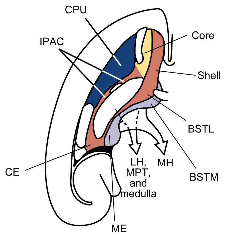
Key areas of the brain (e.g., striatum and extended amygdala) with dopaminergic transition (viewed in a cross-section of the left hemisphere of the brain). The striatum includes the caudate nucleus and putamen (CPU) (dark blue) as well as the nucleus accumbens core (yellow) and shell (red). The extended amygdala is distinguished into a central division (red) and a medial division (light blue). The central division includes the nucleus accumbens shell, the lateral part of the bed nucleus of stria terminalis (BSTL), the central amygdala (CE), and other neuronal groups bridging these areas (IPAC). From these structures, neurons extend to the lateral hypothalamus (LH), visceral nuclei in the brain stem (MPT), and medulla. The medial division of the extended amygdala includes the medial part of the bed nucleus of stria terminalis (BSTM), the medial amygdala (ME), and other associated neuronal groups. Neurons originating in the medial division extend to the medial hypothalamus (MH).
SOURCE: Adapted with permission from Heimer et al. 1997.