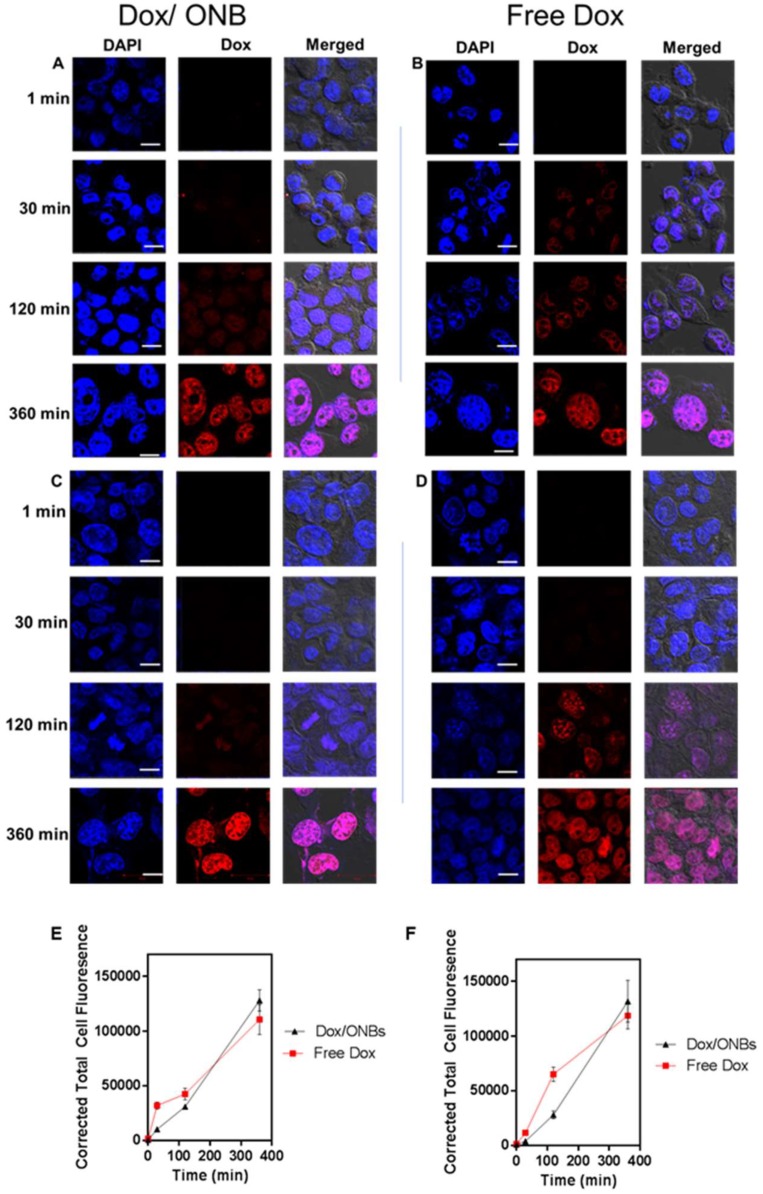
Figure 4.
Intracellular confocal microscopy imaging of Dox penetration using Dox fluorescence and DAPI staining. Dox penetration in MDA-MB-231 cells using (A) Dox/ONB conjugates and (B) Free Dox. Scale bars = 10 µm. Dox penetration in HeLa cells using (C) Dox/ONB and (D) Free Dox. Scale bars = 10 µm. (E) Corrected total cell fluorescence intensity of Free Dox and Dox/ONBs for MDA-MB-231 cells from confocal images measured using ImageJ software. (F) Corrected total cell fluorescence intensity of free Dox and Dox/ONBs for HeLa cells from confocal images measured using ImageJ software.

