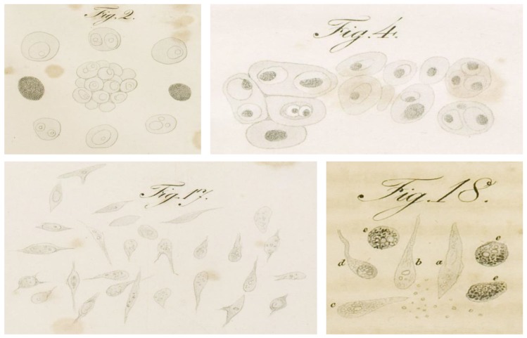
Figure 1.
Reproduction of original drawings representing the microscopic appearance of cancer cells isolated from different human neoplasia, as reported in Muller’s 1838 work on cancer [1]. Panel 2 and panel 17 depict mono- and poly-nucleated tumor cells in a “reticular” carcinoma, and heterogeneous spindle-shaped cells isolated from a lower jaw osteocarcinoma respectively (Table II, page 69); panel 4 represents different polynucleated cells isolated from a tumor of the parotid gland (Table III, page 71); panel 18 shows different morphological cells comprehending pigmented cells (e) isolated from an osteocarcinoma (Table I, page 67).

