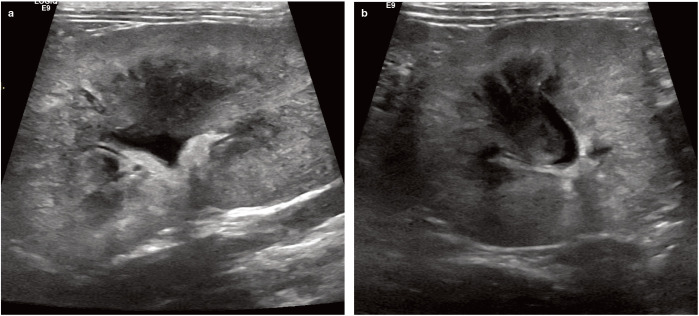
Figure 1.
Longitudinal (a) and transverse (b) ultrasound images of the left kidney of a cat with bilateral renomegaly and acute (grade V) kidney injury. The kidney is enlarged, there is poor corticomedullary differentiation, the cortex is patchy hyperechoic and the medulla has lost its typical hypoechogenic to anechoic appearance. The renal pelvis is dilated to 4 mm. Post-mortem examination of this cat revealed severe pyelonephritis

