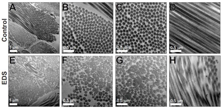
Figure 7.
Structurally abnormal dermal collagen fibrils in Case 1. Transmission electron micrographs from the dermis of control (A–D) and Case 1 (EDS) (E–H). Control dermis contains well-organized collagen fibrils of uniform size and circular profile, while the EDS dermis contains two populations of fibrils. One fibril population is similar to the control dermis, whereas the other heterogeneous population contains large and structurally abnormal fibrils. Scale bars of 0.5 µm or 1 µm are shown.

