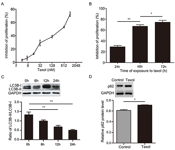
Figure 1. A, Cell proliferation was determined by the CCK-8 assay after treatment with different concentrations of Taxol for 24 h. B, Cell proliferation was determined by the CCK-8 assay after treatment with 31.2 nM Taxol at different time-points. C, Expression of LC3B-I and LC3B-II in MCF-7 cells was determined by western blotting after treatment with 31.2 nM Taxol at different time-points. D, p62 expression in MCF-7 cells was determined by western blotting after treatment with 31.2 nM Taxol for 24 h. Data are reported as means±SD of three independent experiments. *P<0.05, **P<0.01, vs control group (ANOVA).

