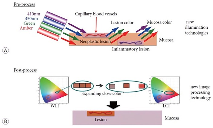
Figure 1.
(a) Pre-processing by linked color imaging (LCI) technology. In neoplastic lesions, the capillaries are located in the shallow layer. 410 nm violet light is absorbed by capillaries, therefore, the violet light is not visible and the neoplastic lesion appears red. In inflammatory lesions, capillaries are in the deep layer. 410 nm violet light is not absorbed, and the lesion appears violet. (b) Post-processing by LCI technology. The colors obtained are separated and reallocated for color enhancement. This makes red and white lesions become more red and more white, respectively.3
WLI: white light imaging.

