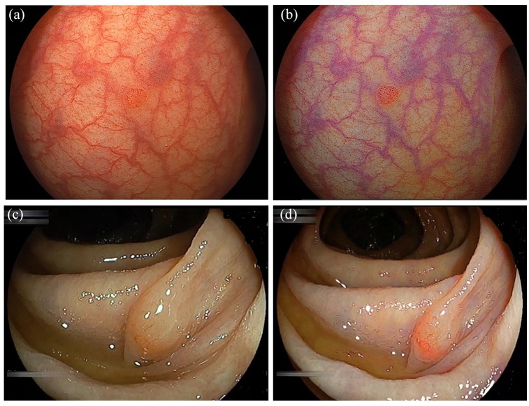
Figure 3.
Detection of colorectal neoplastic lesions. (a) A small polyp in the sigmoid colon seen using white light imaging (WLI). (b) Linked color imaging (LCI) provides clear visualization of the small polyp by enhancing the color contrast. (c) A sessile serrated adenoma/polyp (SSA/P) of the ascending colon seen using WLI. (d) LCI demonstrates a clear line of demarcation by enhancing the color contrast.

