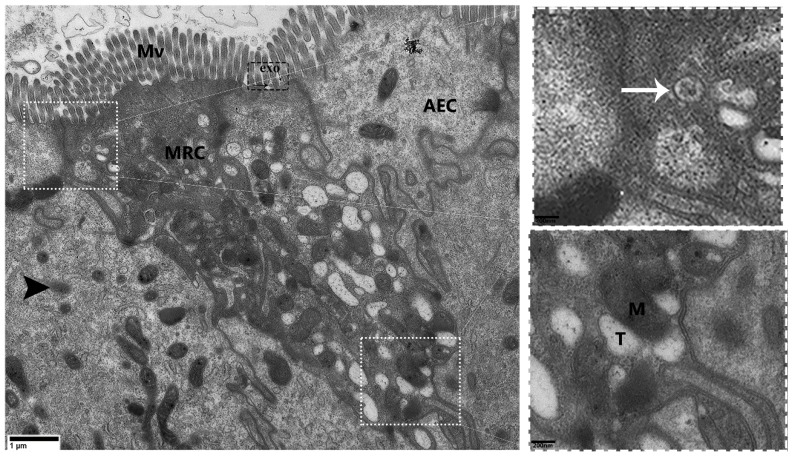
Figure 3.
Transmission electron micrograph of an MRC that exhibits exosome secretion in the mucosal layer of P. sinensis. Mitochondria-rich cells (MRCs), absorptive epithelial cell (AEC), mitochondria (M), tubular system (T), multivesicular bodies (white arrow), exosome (exo), lysosome (black arrowhead), and microvilli (Mv). Scale bar =1 µm.

