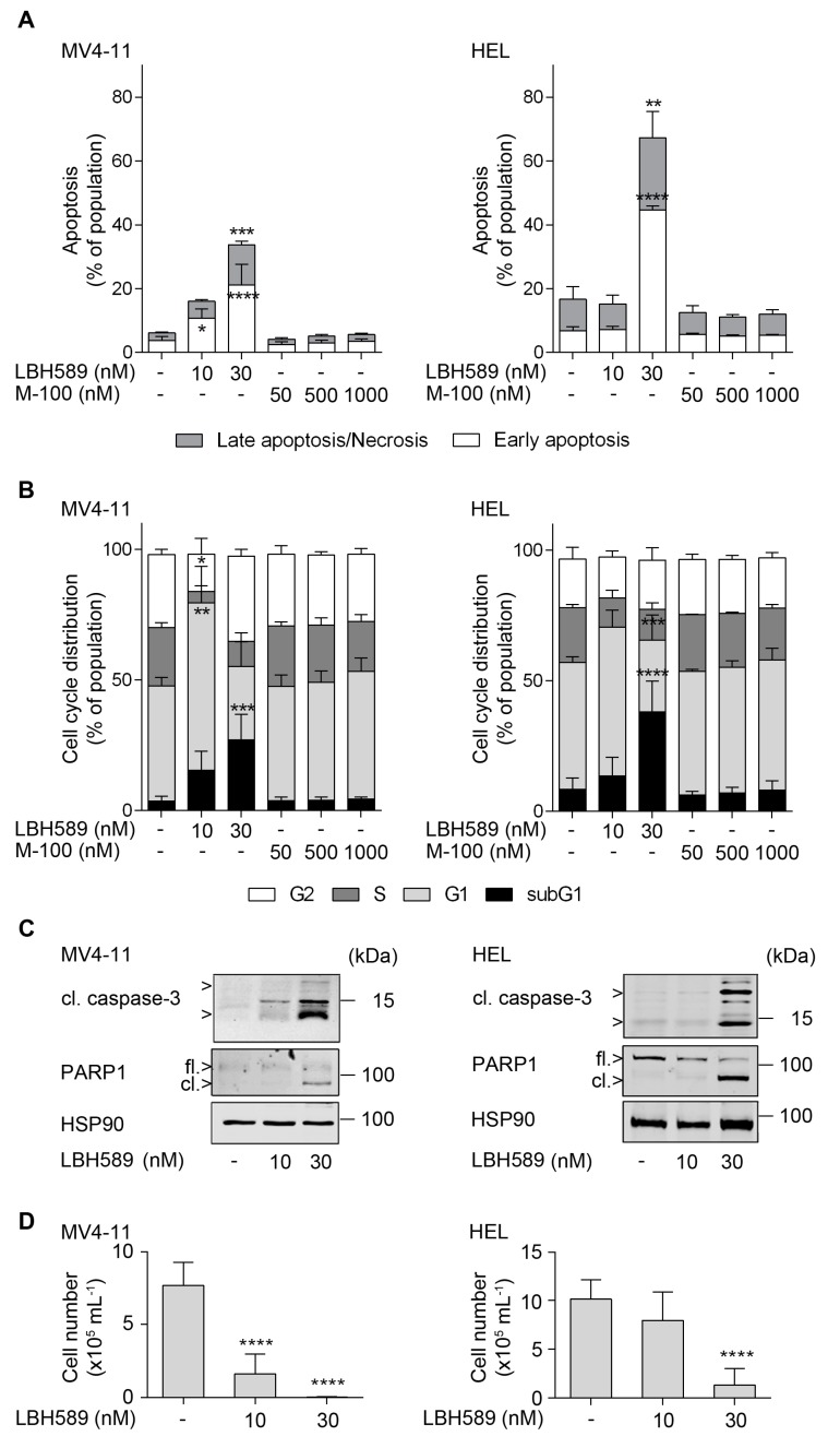
Figure 1.
Human leukemia cell lines are sensitive against LBH589. (A) Human MV4-11 and HEL cells were treated with 10 to 30 nM LBH589 or increasing doses of marbostat-100 (M-100; 50 nM, 500 nM, and 1 µM) for 24 h. Cells were stained with annexin-V-FITC/propidium iodide (PI) and analyzed by flow cytometry (n = 3; mean + SD; two-way ANOVA; * p ≤ 0.05; ** p ≤ 0.01; *** p ≤ 0.001; **** p ≤ 0.0001). (B) Cells were incubated with HDACi as stated in (A). Fixed cells were stained with PI and cell cycle distributions were analyzed by flow cytometry (n = 3; mean + SD; two-way ANOVA; * p ≤ 0.05; ** p ≤ 0.01; *** p ≤ 0.001; **** p ≤ 0.0001). (C) Cells were treated with 10 nM or 30 nM LBH589 for 24 h. Indicated proteins were detected via Western blot (cl., cleaved; fl., full-length) with HSP90 and β-actin as loading controls (n = 3). Please note that compared to HEL cells MV4-11 cells have far less full-length PARP1 and that the lot of the anti-PARP1 antibody may have preferentially recognized the cleaved form of PARP1. (D) Regrowth of the human leukemia cell lines MV4-11 and HEL. Cells were treated with 10 nM or 30 nM LBH589 for 24 h. Thereafter, cells were washed twice with PBS and reseeded. Cells were stained with trypan blue and viable cells were counted after 4 days (n = 3; mean + SD; one-way ANOVA; **** p ≤ 0.0001).

