Figure 3.
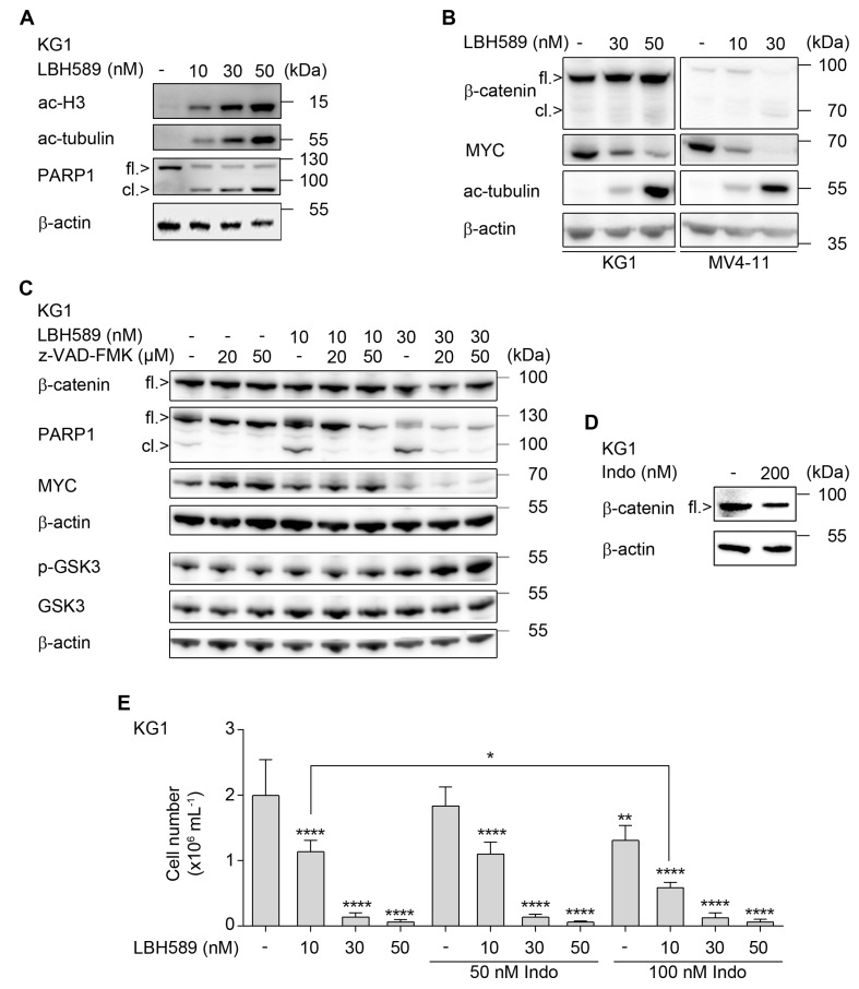
Effects of LBH589 and indomethacin on β-catenin and cell proliferation. (A) KG1 cells were incubated with 10 nM, 30 nM, or 50 nM of LBH589 for 24 h. Indicated proteins were detected via Western blot (fl., full-length; cl., cleaved); with β-actin loading as control (n = 2). (B) KG1 and MV4-11 cells were treated with LBH589 as indicated for 24 h. Western blot was performed for β-catenin, myelocytomatosis oncogene (MYC), acetylated (ac) tubulin, and β-actin as loading control (n = 2). (C) KG1 cells were incubated with 10–30 nM LBH589 ± 20–50 µM z-VAD-FMK for 24 h. Lysates of these cells were analyzed for β-catenin, full-length/cleaved PARP1 (fl./cl.), and MYC. A separate membrane was incubated with antibodies against total and phosphorylated (p) GSK3β; β-actin was used as loading control (n = 2). (D) KG1 cells were cultured in the presence or absence of 200 nM indomethacin for 24 h. Expression of β-catenin was assessed by Western blot; with β-actin as loading control (n = 2). (E) KG1 cells were treated with LBH589 and/or indomethacin as indicated. After 24 h of treatment, substances were washed out and cells were further cultured for proliferation analysis. After 4 days, proliferation was determined by trypan blue exclusion assay. Data are shown as mean values of cells per mL + SD (n = 3; one-way ANOVA; * p ≤ 0.05; ** p ≤ 0.01; **** p ≤ 0.0001).

