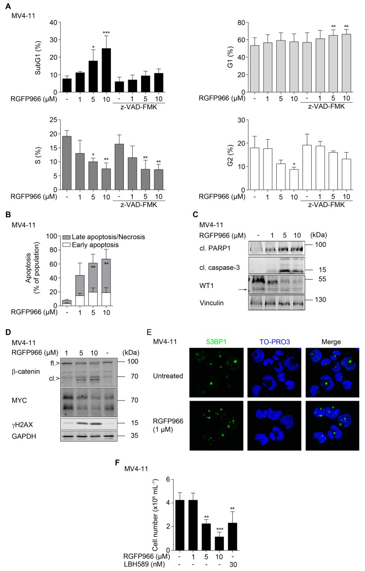
Figure 4.
Biological consequences of HDAC3 inhibition and interplay between β-catenin, MYC, and WT1. (A) MV4-11 cells were pretreated with z-VAD-FMK for 1 h, followed by incubation with RGFP966 as indicated for 24 h. Cells were fixed and stained with PI and analyzed by flow cytometry (n = 3; mean + SD; two-way ANOVA; * p ≤ 0.05; ** p ≤ 0.01; *** p ≤ 0.001). (B) MV4-11 cells were treated as indicated for 24 h. After incubation time, cells were stained with annexin-V-FITC/PI and apoptosis was determined by flow cytometry (n = 3; mean + SD; two-way ANOVA; ** p ≤ 0.01). (C) MV4-11 cells were treated with increasing doses of RGFP966 (1 µM, 5 µM, and 10 µM) for 24 h. Caspase-3, PARP1, and WT1 (→ marks WT1 cleavage product) were analyzed by Western blot, with vinculin as loading control (n = 2). (D) MV4-11 cells were treated as stated in (C). Indicated proteins were detected via Western blot, with GAPDH as loading control (n = 2). (E) Immunofluorescence for 53BP1 in MV4-11 cells that remained untreated or exposed to 1 µM RGFP966 for 24 h. (F) MV4-11 cells were treated as indicated for 24 h. Thereafter, cells were harvested and washed with PBS and reseeded. After 4 days, the cell numbers were determined by trypan blue staining and counting (n = 3; mean + SD; one-way ANOVA; ** p ≤ 0.01; *** p ≤ 0.001).

