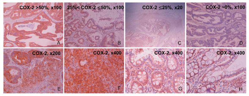
Figure 1.
Representative slides of COX-2 staining in colorectal cancer (CRC) by immunohistochemistry (IHC). Colorectal adenocarcinomas with highly positive COX-2 immunoreaction (>50% of epithelial cancer cells; 22/49 (44.8%) CRC samples) (A), moderately positive (>25% and <50% of epithelial cancer cells; 14/49 (28.6%) CRC samples) (B), mildly positive (<25% of epithelial cancer cells; 5/49 (10.2%) CRC samples) (C), and negative COX-2 immunoreaction (9/49 (18.4%) CRC samples) (D). CRC slides were also imaged at the tumor’s stroma, containing mast cells, fibroblasts, pericytes, and endothelial cells. COX-2 positive stroma staining at ×200 (E) and ×400 (F). Adjacent to the tumor, normal slides were also imaged at the stroma region, with representative slides of COX-2 positive stroma staining at ×400 (G,H).

