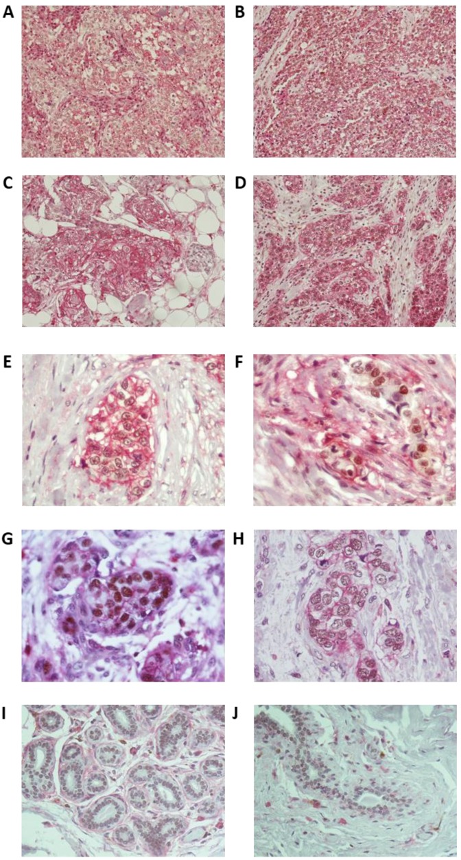
Figure 3.
Immunohistochemistry in breast tumor sections from triple-negative breast cancer (TNBC) patients for LSD1 and the stemness marker CD44. (A–D) Representative images from four different tumors (cases 4, 8, 9, and 10 in Table S1) showing high enrichment in CD44+ cells and high co-expression of CD44 and LSD1. Magnification 200×. (E) Very high (−100%) and (F) lower (−40%) concomitant expression of LSD1 and CD44 in neoplastic cells. (G) Strong and (H) moderate nuclear staining for LSD1 in neoplastic cells. Magnification 600×. (I,J) Non-neoplastic cells of the adjacent breast parenchyma with weak nuclear LSD1 staining from two different tumors (magnification 400×). The cell surface antigen CD44 exhibits membranous staining (chromogenic substrate: magenta) and LSD1 is nuclear (chromogenic substrate: DAB).

