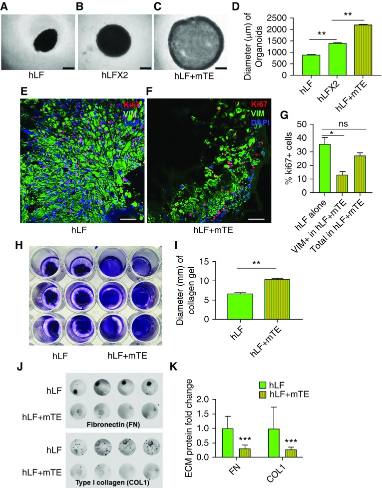
Figure 2.
Organoid culture restrains fibroblast proliferation, contractile function, and extracellular matrix (ECM) deposition. (A) Bright-field microscopy of fibroblasts (1 × 10⁵ hLF) alone in organoid culture conditions. Scale bar: 500 μm. (B) Bright-field microscopy of fibroblasts at twice the seeding density of A (2 × 10⁵ hLF) in organoid culture conditions. Scale bar: 500 μm. (C) Bright-field microscopy of coculture organoids (1 × 10⁵ hLF and 1 × 10⁵ mTEs), with total cell number the same as in B. Scale bar: 500 μm. (D) Quantification of three-dimensional organoid sizes. n = 5. (E–G) Immunofluorescence imaging of Ki-67 staining in human lung fibroblasts (vimentin-positive [VIM+] cells) from organoid-cultured fibroblasts alone (hLF; E) and cocultures (hLF + mTE; F). Scale bar: 50 μm. (G) Quantification of Ki-67+ cells in organoid-cultured fibroblasts alone and cocultures (Vim+ fibroblasts and total cells). n = 3. (H) Collagen gel contraction assay with cells labeled using trypan blue staining. (I) Quantification of collagen gel size showing greater gel compaction by fibroblasts alone and reduced compaction in the presence of mTEs. n = 6. (**P < 0.01). (J) Immunodetection of deposited fibronectin and collagen I in organoid cultures of fibroblasts alone (hLF) and cocultures (hLF + mTE). (K) Quantification of ECM deposition of fibronectin and collagen I. n = 8. *P < 0.05, **P < 0.01, and ***P < 0.001.

