Figure 1.
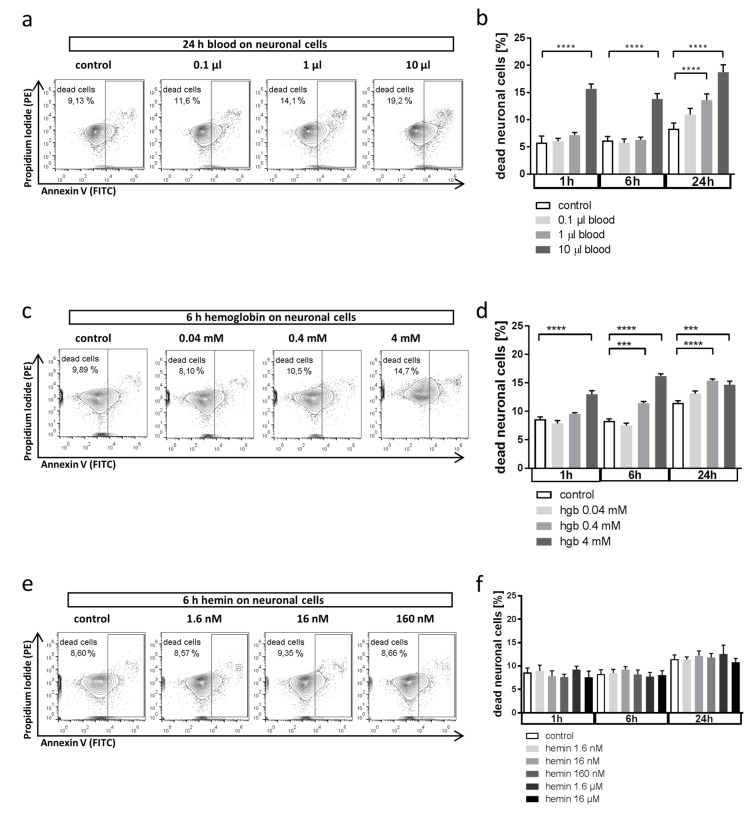
Effect of whole blood and erythrocyte components on neuronal cell death. (a,b) Effect of whole blood incubation on neuronal cell death in HT22 cells in vitro analyzed by flow cytometry after annexin V/PI staining. Cells were exposed to blood at indicated amounts (in 5 mL cell culture medium) and durations before analysis ((a) representative flow cytometry plots; propidium iodide (PI) was detected in the PE channel and annexin V in the FITC channel; (b) quantification as % dead cells from total of n = 6 experiments). **** p < 0.0001 control vs. 10 µL blood 1 h, **** p < 0.0001 control vs. 10 µL blood 6 h, **** p < 0.0001 control vs. 1 µL 24 h, **** p < 0.0001 control vs. 10 µL 24 h. (c,d) Effect of hemoglobin exposure on neuronal cell death in HT22 cells in vitro analyzed by flow cytometry after annexin V/PI staining. Cells were exposed to hemoglobin at indicated concentrations corresponding to the volumes of blood used in (a,b) ((c) representative flow cytometry plots; (d) quantification as % dead cells from total of n = 6 experiments). **** p < 0.0001 control vs 4 mM hemoglobin 1 h, *** p = 0.0002 control vs. 0.4 mM hemoglobin 6 h, **** p < 0.0001 control vs 4 mM hemoglobin 6 h, **** p < 0.0001 control vs. 0.4 mM hemoglobin 24 h, *** p = 0.0002 control vs 4 mM hemoglobin 24 h. (e,f) Effect of hemin on neuronal cell death in HT22 cells in vitro analyzed by flow cytometry after annexin V/PI staining. Cells were exposed to hemin at indicated concentrations corresponding to the volumes of blood used in (a,b) ((e) representative flow cytometry plots; (f) quantification as % dead cells from total of n = 6 experiments). p = n.s. for all comparisons.

