Figure 3.
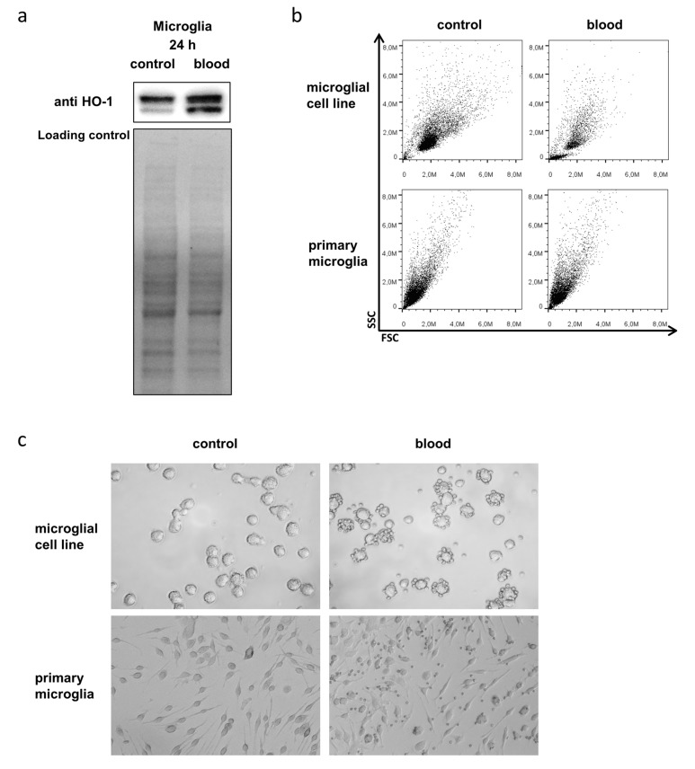
Microglial HO-1 expression and viability in response to blood exposure. (a) Representative Western blotting shows induction of HO-1 protein expression in BV-2 microglia in response to exposure to 10 µL blood for 24 h. Double bands are due to antibody reactivity against the truncated form of HO-1. Lower panel shows corresponding loading control using total protein staining. (b) Representative flow cytometry FSC/SSC dot plots of BV-2 microglia (upper panel) and primary microglia (lower panel) without and with blood exposure (10 µL accordingly). (c) Representative light microscopy images of BV-2 microglia and primary microglia before (left panel) and after blood exposure (right panel; 10 µL accordingly; after washing step to remove erythrocytes). Unchanged morphology, size, and granularity of microglia after exposure to blood indicated unchanged viability.

