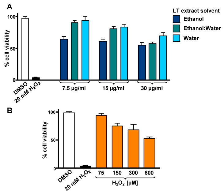
Figure 1.
Cytotoxicity of L. tridentata (LT) extracts (A) and H2O2 (B) on SH-SY5Y cells. The percentage of viable cells was measured using the differential nuclear staining (DNS) assay and a bioimager system. (A) Cells were treated for 24 h with a concentration gradient (7.5, 15 and 30 µg/mL) of each LT extract dissolved with ethanol, ethanol:water (e/w), or water. (B) The percentage of viable cells exposed for 12 h to an H2O2 concentration gradient (75 to 600 µM) was also determined. Dimethyl sulfoxide (DMSO) 0.25% v/v was included as a control. As a positive control for cytotoxicity, 20 mM of H2O2-treated cells were also included. Each bar indicates the average of three biological replicates with its corresponding standard deviation.

