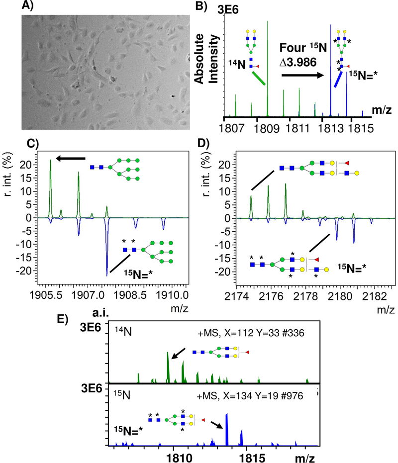
Figure 5.
Detection of stable isotopic labeling in cell culture (SILAC) using Isotopic Detection of Aminosugars With Glutamine (IDAWG) labeling. A) Representative image of human aortic endothelial cells plated at 5,000 cells and cultured for 96 hours with 15N glutamine. 15N incorporates into GlcNac, GalNAc, and sialic acids. B) 15N incorporated into 4 GlcNAc residues of Hex5dhex1HexNac4 bi-antennary N-glycan resulting in a mass shift of 3.986 Da. C) 15N incorporated into 2 GlcNAc residues of Man9, resulting in a 1.9941 Da shift; D) 15N is incorporated into 5 GlcNAc residues of a Hex6dHexHexNAc5 tri-antennary N-glycan resulting in a 4.9852 Da shift. * indicates 15N incorporation. F) Example single spectra from HAEC 14N compared to single spectra of HAEC with 15N labeling. r.int. - relative intensity; a.i. – absolute intensity.

