
Figure 9.
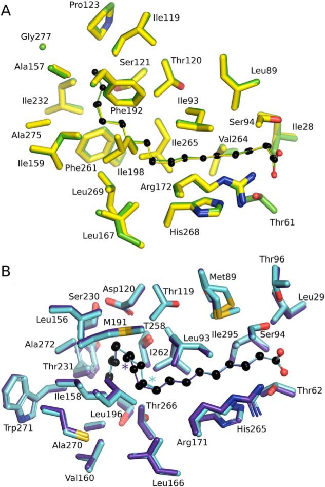
Versatility of FA binding by SpFakB1 and SpFakB2. A, structural alignment of SpFakB1(14:0) (yellow, PDB ID 6NOK) and SpFakB1(16:0) (green, PDB ID 6DKE). The fatty acids are presented as colored sticks with carbon spheres in black. B, structural alignment of SpFakB2 with C18:1Δ9, (cyan, PDB ID 6DJ6) and SpFakB2 with C18:1Δ11 (purple blue, PDB ID, 6NR1). Colored asterisks indicate locations of the sp2 hybridized double bonds. The coordinate alignments were based on the FA using MCSALIGN plugin for PyMOL.