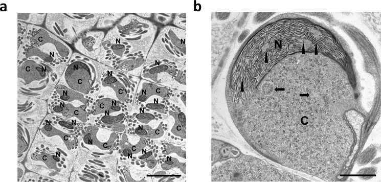
Figure 4.
Electron micrographs of M. polymorpha spermatids. a, chromatin condensing nucleus (N) exhibiting a shrinking cytoplasm (C). The scale bar is 5 μm. b, electron micrograph (greater magnification) of a condensing spermatid showing the characteristic 24 ± 3-nm fibers (arrowheads). The arrows point to autophagosomes. The scale bar is 1 μm.

