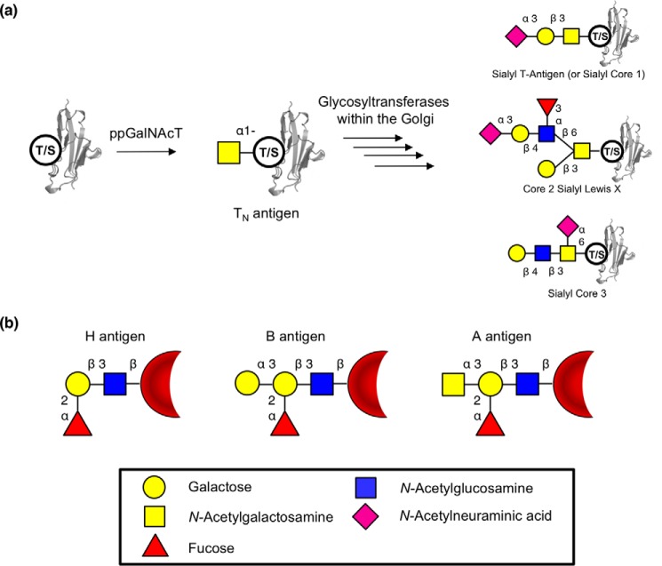
Figure 1.
O-Linked glycans and blood antigens. a, scheme of mucin-type O-linked glycosylation. O-Linked glycans are shown bound to either serine or threonine on the surface of a protein. Structures shown on the right are a selection of known mucin-type O-glycans. b, structures of ABO blood antigens present on red blood cells. All antigens are shown as bound to the surface of red blood cells though a Type 1 linkage. Sugars are shown using the Consortium for Functional Glycomics notation (76).

