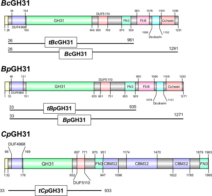
Figure 4.
Protein domain structure of GH31 enzymes in this study. The protein domain structures are presented based on the domains identified based on InterPro prediction (77) and the NCBI conserved domain search (78) and are visualized using DOG2.0 (79). Signal peptides are shown in yellow, whereas the identity and length of all other domains are indicated by color and text within the figure. The truncations of the proteins employed in the present study are displayed below each structure. DUF, domain of unknown function; FN3, fibronectin type 3; F5/8, F5/F8 Type C domain.

