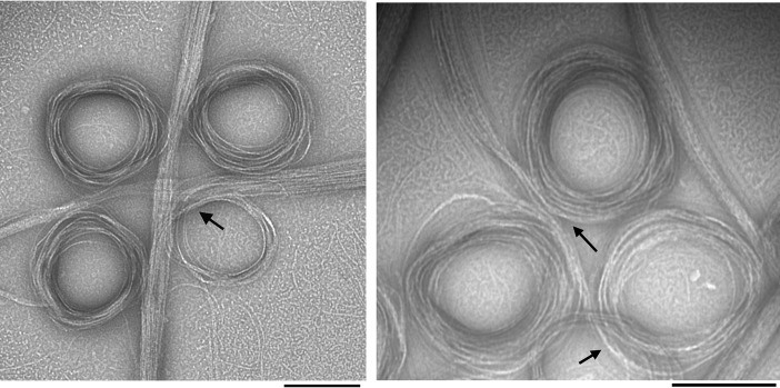
Figure 1.
EM images of negatively stained SyFtsZ pfs after adding 0.5 mm GTP in HMK buffer (50 mm HEPES, 100 mm KAc, 5 mm MgAc) at pH 7.5. 10 μm SyFtsZ assembled into pfs (some isolated pfs are seen in the background), which further associate into straight bundles and toroids. The diameter of the toroids is 200–300 nm. Transitions between straight bundles and toroids are indicated by arrows. Bar, 200 nm.

