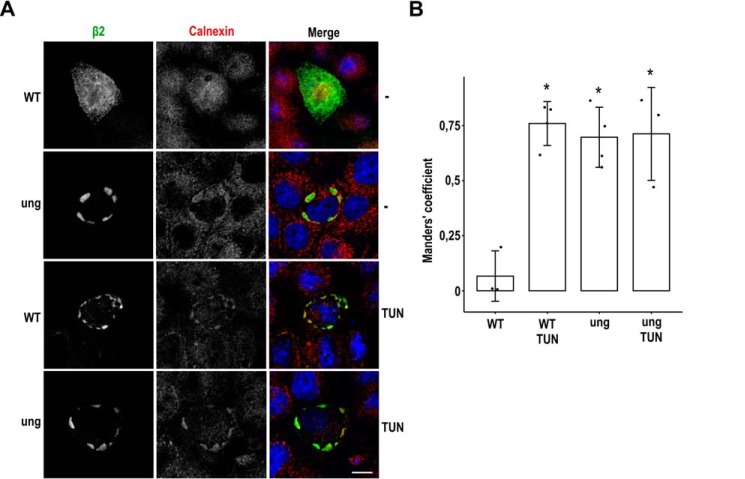
Figure 5.
Unglycosylated β2 is retained in the ER. MDCK cells were transiently transfected with the SCN2B-yfp vector to express WT or fully unglycosylated β2 (ung) and grown for 1 day in wells. Cells were treated with TUN 2 h after transfection or left untreated (−), fixed, and immunostained with a rabbit polyclonal antibody against calnexin (red). A, representative xy sections show that unglycosylated β2 (green) is intracellular and overlaps with calnexin, as does the WT in TUN-treated cells. This contrasts with the localization of β2 WT at the cell end in untreated cells, also displaying a scattered pattern. To focus more accurately where β2 is found in each condition, sections were taken right above the nucleus (WT −) or at the nuclear level (for the rest). Nuclear staining by DAPI is in blue. Scale bar, 10 μm. B, line chart showing Manders' coefficients calculated along the z axis and indicating the fraction of β2 overlapping to compartments labeled with calnexin. The high overlap in TUN-treated cells and in those expressing unglycosylated β2 contrasts with negligible overlap in untreated cells expressing β2 WT. One-way ANOVA with Tukey's HSD post hoc test revealed differences among means (*, p < 0.0005). Data are mean ± S.D. (error bars) (n ≥ 3).

