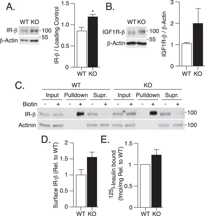
Figure 3.

Attenuated insulin signaling in ATG16L1 KO MEFs is independent of changes at the level of the IR. A, IR-β, and B, IGF1R-β protein content was measured by immunoblotting WT and ATG16L1 KO MEF lysates (n = 3). C and D, cell surface expression of IR-β protein was measured by Sulfo-NHS-SS-Biotin labeling followed by streptavidin pulldown and immunoblotting of input lysates, pulldown and remaining supernatant following pulldown (Supr.) (n = 4). Immunoblots were quantified using Image Studio 5.2.5 software. E, WT and ATG16L1 KO MEFs were treated with 0.5 nm 125I-insulin on ice for 20 min to evaluate insulin binding. After washing away excess 125I-insulin, cells were lysed, radioactivity and protein content were measured, and the fmol/mg of bound 125I-insulin was calculated (n = 3). For IR-β expression, a one-tailed paired t test was performed, as others have shown the IR to be degraded by autophagy. For all other measures, p value was calculated using a two-tailed paired t test. *, p < 0.05.
