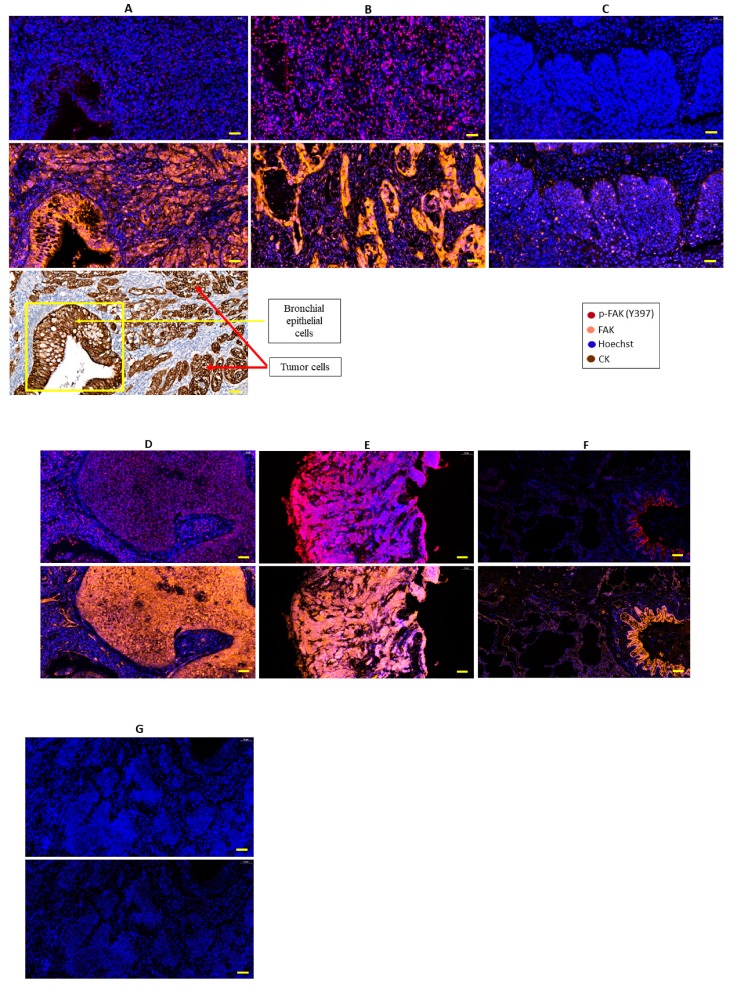
Figure 2.
Illustrations of FAK and phospho-FAK (Y397) expression evaluated by multiplex immunofluorescence (IF) immunohistochemistry (IHC) in lung cancer and normal lung tissues. (A) Lung adenocarcinoma with the absence of phospho-FAK expression but homogenous cytoplasmic FAK staining (orange) in the tumor core, adjacent non-tumoral bronchi, and some stromal cells (including vessels and lymphoid structures). (B) Lung adenocarcinoma with nuclear phospho-FAK staining (red) and homogenous cytoplasmic FAK staining (orange). (C) Lung squamous carcinoma with the absence of phospho-FAK expression but weak cytoplasmic FAK staining. (D) Lung squamous carcinoma with nuclear phospho-FAK staining (red) and homogenous cytoplasmic FAK staining (orange). (E) Small-cell lung cancer with nuclear phospho-FAK staining (red) and cytoplasmic FAK staining (orange). (F) Normal lung with cytoplasmic FAK staining in bronchi and some stromal cells (including vessels and lymphoid structures). (G) Lung squamous carcinoma used as a negative control, showing the absence of phospho-FAK and FAK staining. Original magnification: 20×; scale bar: 50 µm.

