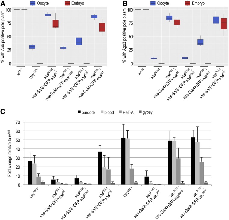
Figure 3.
Localization of Aub and Ago3 in the egg chamber and embryos depends on Vasa. (A) Box plot showing percentage of oocytes and embryo progeny of wild-type (w1118), vasPD/D1, vasPD/D1; vas-Gal4 > GFP-VasWT, vasPD/D1; vas-Gal4 > GFP-VasDQAD, vasPD/D1; vas-Gal4 > GFP-VasGNT, and vasD1/D1; vas-Gal4 > GFP-VasWT flies displaying Aub-positive pole plasm, as determined by immunohistochemical detection of Aub. Experiments were performed in three independent replicates. (B) Box plot representing percentage of oocytes and embryo progeny of wild-type (w1118), vasPD/D1, vasPD/D1; vas-Gal4 > GFP-VasWT, vasPD/D1; vas-Gal4 > GFP-VasDQAD, vasPD/D1; vas-Gal4 > GFP-VasGNT, and vasD1/D1; vas-Gal4 > GFP-VasWT flies displaying Ago3-positive pole plasm, as determined by immunohistochemical detection of Ago3 protein. Experiments were performed in three independent replicates. (C) Quantitative PCR analysis for LTR transposons burdock, blood, and gypsy and non-LTR transposon HeT-A in vasPD/D1, vasPD/D1; vas-Gal4 > GFP-VasWT, vasPD/D1; vas-Gal4 > GFP-VasDQAD, vasPD/D1; vas-Gal4 > GFP-VasGNT, vasPD/D1, vasPD/D1; vas-Gal4 > GFP-VasWT, vasPD/D1; vas-Gal4 > GFP-VasDQAD, and vasPD/D1; vas-Gal4 > GFP-VasGNT flies. Expression of transposons in wild-type (w1118) was set to 1 and normalized to rp49 mRNA in individual experiments. Error bars represent SD from three biological replicates.

