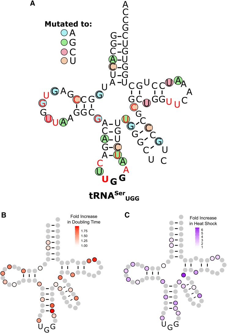
Figure 4.
Randomly selected second-site mutations that dampen mistranslation by tRNASerUGG. (A) Base changes are indicated by the colored circles, where blue indicates a mutation to adenine, green to guanine, red to cytosine, and orange to uracil. Bases colored in red are sites of modification in tRNASer (Machnicka et al. 2013). (B) Heat map of the growth rates of strains containing mutations in tRNASerUGG that mistranslate at a level that supports viability. BY4742 containing tRNASerUGG variants were grown to saturation in media lacking uracil, diluted to an OD600 of ∼0.1 in the same media and grown for 24 hr at 30°. OD600 was measured every 15 min. Doubling time was calculated with the R package “growthcurver” (Sprouffske and Wagner 2016) and normalized to a strain containing a wild-type tRNASer. (C) Heat map of heat shock induced by tRNASerUGG derivatives. BY4742 containing different tRNASerUGG variants and a fluorescence heat shock reporter were grown to saturation in media lacking uracil in biological triplicate. Cells were diluted 1:20 in the same media and grown for 6 hr. Cell densities were normalized and fluorescence measured.

