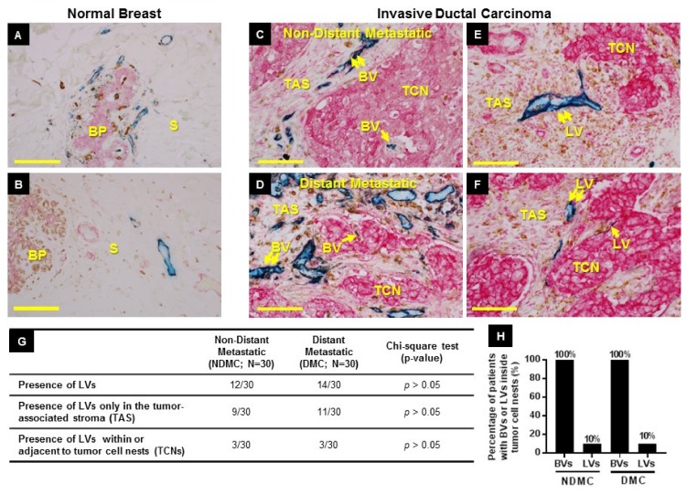
Figure 1.
Location of blood vessels (BVs) and lymphatic vessels (LVs) in normal and neoplastic breast tissue. (A,B) Normal breast. (A) BVs (blue, CD31) are seen in close proximity to the breast glandular parenchyma (BP) as well as in the adjacent connective tissue stroma (S). (B) LVs (blue, D2-40) are seen in the stroma (S). (C–F) BVs and LVs in primary tumors of patients with metastatic and non-metastatic breast cancer. (C,D) In all non-distant metastatic (C) and distant metastatic (D) cases, BVs are found both in tumor cell nests (TCNs) (single arrow) and in tumor-associated stroma (TAS) (double arrow). (E,F) In some cases (E), LVs are found only in TAS (double arrow), and only rarely (F) are LVs found both in TAS (double arrow) and in TCNs (single arrow). Scale bars (A–F) = 90 µm. (G) Frequency of LV presence in the primary tumors of non-distant metastatic and distant metastatic patient cohorts. (H) Percentage (%) of patients with BVs or LVs inside tumor cell nests in the primary tumors of non-distant metastatic and metastatic patient cohort. See Table 2 for panels C–G raw data.

